Services


Optometry Services in Burney, CA
We are committed to providing quality eye care to you and your family.
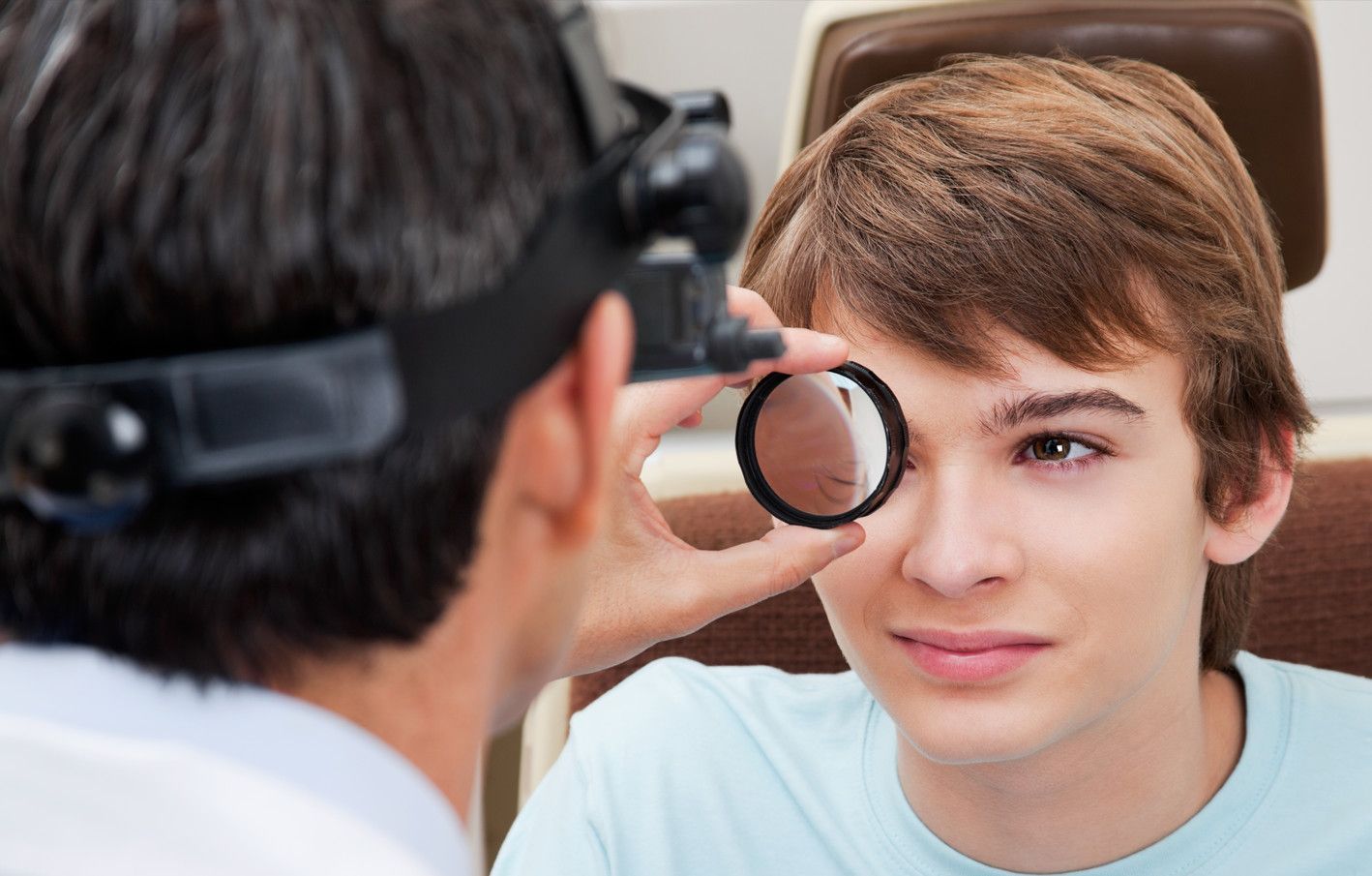
Comprehensive Eye Exam
What Are The Benefits Of A Comprehensive Eye Exam?
Comprehensive eye exams evaluate all aspects of your vision and eye health.
Internal Exam – This is an evaluation of the retina and optic nerve while your eyes are dilated.
Visual Function and Eye Health – This includes testing depth perception, color vision, peripheral vision and response of the pupils to light, as well as an evaluation of eye focusing, eye teaming and eye movement abilities.
Glaucoma Testing – This is a test of fluid pressure within your eyes to check for the possibility of glaucoma.
Visual Acuity – Your doctor will test your vision with different lenses to determine if glasses or contact lenses can improve your vision.
Comprehensive eye exams look at your total health history.
Even though you visit a separate office for your eye health, that doesn’t mean your eyes shouldn’t be treated holistically. Your eye doctor will discuss your overall health and that of your immediate family, any medications you’re taking and whether you have high blood pressure or diabetes. They’ll also want to know if you smoke and how much sun exposure you get. All these factors help the eye doctor properly assess your eye health.
Comprehensive eye exams are performed by eye professionals.
Optometrists are highly trained and will examine the eyes for visual defects, diagnose problems or impairments, and prescribe corrective lenses. After a bachelor's degree, optometrists complete a four-year program to obtain their Doctor of Optometry degree.
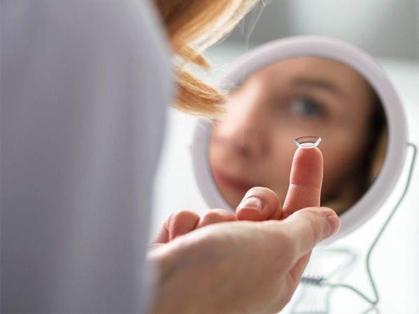
Contact Lenses Exam
If you’ve never worn contact lenses, it can feel a bit intimidating. After all, you’re inserting something into your eye! Let’s ease your mind about the first step – your contact lens exam. This blog will walk you through what’s involved in a contact lens exam and what you can expect every step of the way.
It begins with a comprehensive eye exam.
Your eye doctor will first determine your overall eye health and vision. This includes a discussion of your health history and then a series of standard eye tests. These tests will evaluate eye focusing, eye teaming, depth perception, color vision, peripheral vision, and the response of pupils to light. The doctor will also measure your eye’s fluid pressure to check for glaucoma, evaluate your retina and optic nerve, and test your vision with different lenses to assess whether contact lenses can improve your vision.
Then, a discussion about your contact lens preferences.
If contact lenses are appropriate for you, it’s time to talk about your contact lens preferences. For example, do you want to enhance or change your eye color? Would you prefer daily disposable lenses or overnight contacts? Ask about the benefits or drawbacks of each, so you make the best decision. If you’re over 40, your doctor will likely discuss age-related vision changes and how contact lenses can address these issues.
Next, the eye doctor will conduct eye surface measurements.
Contact lenses require precise measurements of your eyes to fit properly. Using an instrument called a keratometer, your doctor will measure the curvature of your eye's clear front surface. This is your cornea. Next, the size of your eye's pupil is measured using a card or ruler showing different pupil sizes. This is held next to your eye to determine the best match.
You may also need a tear film evaluation.
If you have dry eyes, your eye doctor will perform a tear film evaluation to measure the amount of tear film on the surface of your eye. If your tear film is insufficient or you have chronic dry eyes, contact lenses may not be a good option for you. However, some newer contact lenses deliver moisture to the surface of the eye, making them a better choice for individuals with dry eye issues.
It's time for the contact lens fitting.
The final step is to fit you with a trial pair of contact lenses. Once inserted, your eye doctor will examine the lenses in your eyes to ensure a good fit. He/she will check the alignment and movement of the lenses on the surface of your eye. If the fit looks good, the last step is to ensure the prescription is correct with several tests.
Now it’s your turn to test it out.
Your contact lens exam is over, but you’ll need to come back. Your doctor will usually have you wear the trial lenses for a week. Then you’ll have a short follow-up exam to confirm that the lenses are working well for you. You can then order a supply of contact lenses.
If this is your first contact lens exam, don’t worry. Choose a qualified optometrist and they’ll answer all your questions as you go. Just be sure to let them know you’re interested in contact lenses. That way, they can allow extra time in your appointment for specialized tests and consultation.
Pediatric Eye Exams
Regular eye exams are important for children because their eyes can change significantly in as little as a year as the muscles and tissue development. Good eyesight is critical for a child’s life and achievements; success in school is closely tied to eye health. School demands intense visual involvement, including reading, writing, using computer and blackboard/smartboard work. Even physical activities and sports require a strong vision. If their eyes aren’t up to the task, a child may feel tired, have trouble concentrating, have problems in school and have difficulty playing their favorite games which may affect their quality of life.
When To Perform A Pediatric Eye Exam?
According to the recommendations of the American Academy of Ophthalmology and the American Association for Pediatric Ophthalmology and Strabismus, a child should have initial screening between 6 and 12 months of age followed by routine eye health and vision screenings throughout childhood to help detect any abnormalities as their eyes develop every two years thereafter until the age of 18, unless otherwise recommended.
For a newborn, an optometrist should examine the baby’s eyes and perform a test called “red reflex test” which is a basic indicator that the eyes are normal. In case that, the baby is premature or at high risk for medical problems for other reasons, has signs of abnormalities, or has a family history of serious vision disorders in childhood, the optometrist should perform a comprehensive exam.
A second eye health examination should be done to infants between six months and one year of age. This examination includes tests of the pupil responses to evaluate whether the eyes pupil opens and closes properly in the presence or absence of light, a fixate and follow test to determine whether the baby can fixate on an object, such as light, and follow it as it moves, and a preferential looking test by using cards that are blank on one side with stripes on the other side to attract the gaze of an infant to the stripes and assess the vision capabilities, which infants should be able to perform well by the time they are 3 months old.
For a Preschooler, between the ages of 3 and 3½, a child’s visual acuity and eye alignment should be assessed. If the child is diagnosed with misaligned eyes (strabismus), "lazy eye” (amblyopia), refractive errors (astigmatism, myopia, hyperopia) or any other focusing problems, it’s important to begin treatment as soon as possible to ensure successful vision correction and life-long benefits.
At school age, or upon entering school, the child’s eyes should be screened for visual acuity and alignment. In this age group, nearsightedness (myopia) is the most common refractive error and can be corrected with eyeglasses.
There are some signs that parents can tell if their child has a vision problem, for example, the child may squint, hold reading material very close to face, or complain about things appearing blurry. However, there are some less obvious signs that may indicate vision problems, such as having a short attention span, quickly losing interest in games, projects or activities that require using their eyes for an extended period of time, or losing their place when reading. Also, choosing to avoid reading, drawing, playing games or doing other projects that require focusing up close. Another sign is that a child may turn his or her head to the side when looking at something in front of them. This may be a sign of a refractive error, including astigmatism, so by turning their head helps the child see better.
That’s why it is so important for kids to have regular eye screenings. The earlier a vision problem is found and treated, the better off your child will be in and out of school.
Laser Eye Surgery Co-Management
LASIK (laser-assisted in situ keratomileusis), is the most popular refractive surgical procedure. In this procedure, a laser is used to permanently change the shape of the cornea (the clear covering on the front of the eye) to correct common vision problems such as nearsightedness, farsightedness, astigmatism, and presbyopia. This improves vision and reduces a person's need for glasses or contact lenses.
LASIK uses an excimer laser (an ultraviolet laser) to remove a thin layer of corneal tissue, giving the cornea a new shape, so that light rays are focused clearly on the retina. In the case of a nearsighted person, the goal of LASIK is to flatten the too-steep cornea; with farsighted people, a steeper cornea is desired. LASIK can also correct astigmatism by smoothing an irregular cornea into a more normal shape.
LASIK is an outpatient surgical procedure with no need to stay at the surgery center overnight as it will take 10 to 15 minutes to perform for each eye. The procedure is done while the patient is awake, but the patient may request mild sedation. The only anesthetic used is eye drops that numb the surface of the eye. LASIK can be done on one or both eyes during the same session.
How To Prepare For LASIK Eye Surgery?
Before LASIK eye surgery, the eye surgeon will evaluate the patient’s medical history and perform a full eye examination, including measuring corneal thickness, refraction, corneal mapping, eye pressure, and pupil dilation. Afterward, the surgeon will discuss what to expect during and after the procedure.
On the day of the surgery, eat a light meal before going to the doctor and take all prescribed medications, if any. Do not wear eye makeup, creams, perfumes or lotions on the day before and the day of surgery, or have any bulky hair accessories that will interfere with positioning head under the laser.
Contact lenses shouldn't be worn for at least three days prior to the evaluation. In the case of, rigid gas permeable contact lenses, they should not be worn for at least three weeks before. Patients should arrange for a ride home from the place of surgery, as their vision might be blurry.
What Happens During LASIK Eye Surgery?
The LASIK surgeon uses a computer to adjust the laser for each patient’s particular prescription. An instrument to hold the eyelids open may be used and the patient will be asked to look at a target light for a short time while the laser sends pulses of light to painlessly reshape the cornea. During LASIK eye surgery, a suction ring is placed on the eye just before cutting the corneal flap that may cause a feeling of pressure and may cause vision to dim slightly. Then, an instrument called a femtosecond laser is used to create a thin flap in the cornea. The corneal flap is then painlessly peeled back and the underlying corneal tissue is reshaped using another laser. After the cornea is reshaped so that it can properly focus light onto the retina, the cornea flap is put back in place and the surgery is complete. A distinct odor might be detected as the laser removes the corneal tissue which some people describe as similar to that of burning hair, but is nothing to worry about.
What To Expect After LASIK Eye Surgery?
The eyes might temporarily be dry even though they do not feel that way. One eye drop will be prescribed to prevent infection and inflammation and another eye drop to keep eyes moist. These drops may cause a momentary slight burn or blurring of your vision upon using them. Do not use any eye drops not approved by the LASIK surgeon.
Healing after LASIK eye surgery usually occurs very rapidly. Vision may be blurry and hazy for the first day, but most patients notice improved vision within a few days of surgery. There will be a follow-up evaluation 24 to 48 hours after LASIK eye surgery, as well as at regular intervals within the first six months.
What Are The Advantages Of LASIK Eye Surgery?
LASIK has many benefits, including:
Vision is corrected nearly by the day after LASIK.
LASIK causes a dramatic reduction in eyeglass or contact lens dependence and many patients no longer need them at all.
Adjustments can be made years after LASIK to further correct vision if vision changes with age.
LASIK is associated with very little pain due to the numbing eye drops that are used.
No bandages or stitches are required after LASIK.
Glaucoma Management
You might be surprised at how many tests eye doctors use to diagnose glaucoma. A proper diagnosis requires careful evaluation of many aspects of your eye’s health – from eye pressure to cornea thickness to the health of your optic nerve. This article describes how your eye doctor assesses your risk and all the tests needed to properly diagnose glaucoma.
Risk Factor Assessment
Your eye doctor will begin by assessing your risk level for developing glaucoma. This will help determine the frequency and extent of testing needed. Through a family history and medical questionnaire, the eye doctor is looking for the following risk factors:
Over the age of 60
Ethnic background such as African or black Caribbean descent, Hispanic, or Asian
Family history of glaucoma, such as a sibling or parent with glaucoma
History of eye conditions, injuries or surgeries
Prolonged corticosteroid use (eye drops, pills, inhalers or creams)
Chronic conditions that affect blood flow, such as migraines, diabetes, low blood
pressure or hypertensionCurrent or former smoker
If you’ve already had a comprehensive eye exam, your eye doctor will also consider these risk factors:
Eye pressure higher than normal (above 21 mm Hg)
Thin corneas (less than 0.5 millimeters)
Your type of eyesight is also important. People with farsightedness are at a higher risk for narrow-angle glaucoma, a more serious type that can advance quickly. While nearsightedness is associated with open-angle glaucoma, which progresses slowly without any symptoms.
Standard Glaucoma Tests
During a comprehensive eye exam, your eye doctor will always check for glaucoma, regardless of the risk level. This provides a baseline for comparison as you age. There are two tests: tonometry and ophthalmoscopy.
Tonometry
Tonometry measures the pressure within your eye. Your eye doctor will give you drops to numb your eyes. Then he/she will use a device called a tonometer, which applies a small amount of pressure with a warm puff of air.
Eye pressure is unique to each person, so it’s not always a reliable indicator for glaucoma. It’s simply another piece of information to help your eye doctor assess your eyes. The range for normal pressure is 12-22 mm Hg (“mm Hg” in millimeters of mercury, a scale for recording eye pressure). Most glaucoma cases are diagnosed with pressure over 20mm Hg. However, some people can have glaucoma at pressures between 12 -22mm Hg.
Ophthalmoscopy
This is an examination of your optic nerve. Your eye doctor will use eye drops to dilate the pupil, which makes it possible to see through your eye to examine the shape and color of the optic nerve. Then, using a small device with a light on the end, your optic nerve is magnified. Based on the results of these tests, your doctor may ask you to have more glaucoma exams.
Supplemental Glaucoma Tests
Perimetry
Perimetry is a visual field test. It creates a map of your complete field of vision. During this test, you’ll look straight ahead and then indicate when a moving light passes your peripheral (or side) vision. Try to relax and respond as accurately as possible. To ensure accuracy, your doctor may repeat the test to see if the results are the same the next time. If you’ve been diagnosed with glaucoma, a visual field test is usually recommended at least once per year to assess changes to your vision.
Gonioscopy
This diagnostic exam helps determine the angle of your iris and cornea. First, you’ll receive eye drops to numb the eye. A hand-held contact lens is gently placed on the eye. A mirror on the contact lens shows the doctor if the angle is closed and blocked (a possible sign of angle-closure or acute glaucoma) or wide and open (a possible sign of open-angle, chronic glaucoma).
Pachymetry
Last, your eye doctor may want to use pachymetry as another way to confirm a diagnosis. Pachymetry measures the thickness of your cornea, the clear window at the front of the eye. A probe called a pachymeter is gently placed on your cornea to measure its thickness. Pachymetry can help clarify your diagnosis because corneal thickness has the potential to influence eye pressure readings.
Glaucoma diagnosis is not as simple as you might expect. Be sure to have regular eye exams, especially if you have any of the risk factors, to detect glaucoma early.
Diabetic Exams
You have almost certainly heard of diabetes, which is one of the most common chronic health conditions in the United States with an estimated 100 million adults currently living with diabetes or pre-diabetes. This metabolic disorder occurs when the body is no longer able to regulate its own blood sugar levels and requires intervention to keep them stable. Most people are aware that diabetes can have serious consequences for our health. However, you may be surprised to learn that it can also influence our vision. This is because patients who are diabetic can go on to develop a complication that is known as diabetic retinopathy. Without prompt treatment, diabetic retinopathy can cause permanent vision loss. It is for this reason that patients who suffer from diabetes are asked to attend regular diabetic-related eye exams.
What Is Diabetic Retinopathy?
For us to be able to see clearly, our eyes need to be healthy and functioning perfectly. The most important component of our eyes are the retina. Found at the very back of the eye, the retina is a patch of light-sensitive cells that have the job of converting the light that passes into the eye into messages that are passed up the optic nerve and into our brain. Our brain then receives them and tells us what we can see and how clearly we can see it.
The retina relies on a continuous supply of blood, which is delivered using a network of tiny blood vessels. Over time, having continuously high blood vessels can damage these blood vessels causing a leak of blood and other fluids onto the retina. If this happens, scarring may occur which could compromise the quality of your vision.
Am I At Risk Of Diabetic Retinopathy?
Technically, anyone who suffers from diabetes, whether it be Type 1 or Type 2, could be at risk of developing diabetic retinopathy. However, the condition is more likely in certain situations. These include if:
- your blood sugar levels are uncontrolled or poorly controlled
- you have a long history of diabetes
- you have high blood pressure (hypertension)
- you suffer from high cholesterol
- you are pregnant
Regular diabetic-related eye exams will enable your eye doctor to monitor your condition and ensure that any signs of diabetic retinopathy are detected and acted upon immediately.
What To Expect From Diabetic-Related Eye Exams?
The process of a diabetic eye exam is very simple and straightforward. In fact, in most instances, it is included within the other elements of comprehensive eye exam and you may not even realize that you have had a specific test to check for diabetes-related complications.
Diabetic eye screening is non-invasive. You will be given eyedrops which will blur your vision. These may sting a little when they are administered, but this will pass within just a few moments. Once your vision is blurred, you will be asked to rest your head onto a device and stare down a lens. This leads to a camera which will take images of the backs of your eyes so that your eye doctor can assess the structures, which include the retina, for any abnormalities. You will see a flash when each image is taken, but at no point should you be in any pain.
In addition to the images of the back of your eye being taken, you will also be given a visual acuity test. This is where you will be asked to read letters off a chart a short distance away, as well as reading from a card held in front of you.
The information that your eye doctor will obtain from your examination will be able to tell them if you are experiencing any of the signs of diabetic retinopathy. If so, they will discuss the best way to get your condition under control. This could involve a combination of elements, including controlling your diabetes more effectively, taking medications or more invasive treatment to preserve your vision. Your eye doctor will give you more specific information based on your individual circumstances.
If you have further questions about diabetic-related eye exams, please contact our knowledgeable eye care team.
Macular Degeneration
Macular degeneration, commonly referred to as age-related macular degeneration (AMD), is the single largest cause of sight loss in the developed world and affects more than 10 million Americans. It usually affects people over the age of 60, but has been known to affect those who are younger. It is a painless condition that usually affects both eyes with the loss being experienced in the central vision. It does not affect the peripheral vision, meaning that it does not cause total blindness.
Varieties Of AMD
Wet AMD
Wet AMD is one variety of the condition in which abnormal blood vessels grow into the macula, leaking blood or fluid which then causes scarring and a rapid loss of central vision. Wet AMD can develop suddenly and rapid referral to a specialist is essential as it can be treated if caught quickly.
Dry AMD
Dry AMD is the most common variety of age-related macular degeneration and is a gradual deterioration of the retina as the cells die off over time and are not regenerated. Up to 15% of people with dry AMD go on to develop wet AMD, and so any sudden changes in your vision should be followed up with your optometrist as soon as possible.
Symptoms Of Macular Degeneration
Macular degeneration affects each person differently, which means that it can sometimes be difficult to diagnose, particularly as you may not notice any change in your vision early on in the condition. However, as the cells deteriorate, you will start to see an increasing range of symptoms, including:
Distortion or bends in what should be straight lines (such as lampposts or door frames)
Dark spots in your central vision
Fading colors
Difficulty adapting from dark to light environments
Blurred vision
Objects may appear to change shape, size or color, or may move or disappear
Bright lights may be difficult to tolerate
Words may disappear while you are reading
Is There Any Way I Can Reduce My Risk Of Developing AMD?
Unfortunately, there is no clear reason as to what triggers the process that causes macular degeneration. However, you are at an increased risk if you have a family history of the condition, or if you are over 60.
Experts suggest that the best thing you can do to minimize any potential risk is to ensure that you live a healthy, active lifestyle. You can do this by:
Stopping smoking
Eating a healthy, balanced diet with plenty of fruit and vegetables
Moderating your alcohol consumption
Maintaining a healthy weight
Getting regular exercise
There is also some limited research that suggests that eating leafy, green vegetables can slow the deterioration of vision in cases of dry AMD.
Treatment For AMD
Sadly, there is currently no cure for either variety of AMD. In the case of dry AMD, the treatments suggested are done so with the aim of aiding the patient to make the most of their remaining vision. This can include things such as using magnifying glasses to help with reading.
Wet AMD can be treated with anti-vascular endothelial growth factor medication. This should stop additional blood vessels from developing and stop your vision from deteriorating further.
Occasionally, laser therapy is suggested as a possible treatment for destroying abnormal blood cells, but this is only suitable for cases of wet AMD and usually only around 1 in 7 sufferers may be potential candidates for this procedure.
If you have any questions or concerns regarding macular degeneration, we highly recommend that you speak with your optometrist who will be happy to assist you.
Cataracts
If you’ve been diagnosed with cataracts, you may wonder if cataract surgery is right around the corner. Not to worry. There are many preventive steps you can take to slow the progression of cataracts and preserve your vision. That doesn’t mean you won’t eventually need surgery, but you can at least delay the need for quite a while.
Protect Your Eyes From The Sun
The National Eye Institute recommends protecting your eyes from the sun's harmful ultraviolet (UV) and high-energy visible (HEV) rays by always wearing good quality sunglasses while outdoors. Look for sunglasses that block 100 percent of UV rays and absorb most HEV rays with large lenses or a close-fitting wraparound style. Remember that the peak hours for sun exposure are between 10 am and 3 pm or 11 am and 4 pm during daylight savings time and that the sun’s rays are strong enough to pass through clouds, so you need your sunglasses every day.
Avoid Steroid Eye Drops
Steroid eye drops are routinely prescribed to treat dry eyes or an arthritic flare-up in the eyes. Unfortunately, they can also speed up the progression of cataracts. Talk to your Optometrist about how you can manage both conditions without inadvertently making your cataracts worse – and hastening the need for surgery.
Check Your Medications
There are over 300 commonly prescribed medications with side effects that may impact cataract progression. Since your primary care physician may not have access to your eye doctor’s medical records, be sure to ask your doctor if your current medications will affect your cataracts. If you must stay on the medication, it’s even more important to avoid sunlight during peak hours and to wear sunglasses.
Quit Smoking
If you haven’t quit already, here’s another good reason to do it: over time, the damage from smoking can double or triple an individual's risk of developing cataracts. If you’ve been a smoker, your habit was probably a big contributor to the diagnosis. The good news is – by quitting smoking now, you can slow the progression of cataracts.
Follow Eye Health Diet Guidelines
Studies have shown that certain vitamins and nutrients may reduce age-related decline in eye health, particularly antioxidants. If you’ve already been diagnosed with cataracts, adding foods rich in antioxidants to your diet will help slow the progression. This list isn’t exhaustive, but here are some examples to get you started: dark chocolate, blueberries, strawberries, pecans, carrots, sweet potatoes, artichokes, kale, red cabbage, beans, beets, spinach, apples, and plums.
Doctors also recommend eating more fish high in omega-3 fatty acids. This has been linked to a potentially reduced risk of cataracts or their progression. You may also consider taking a multivitamin that contains Vitamin C and E. Talk to your doctor or nutritionist about how you might adopt a healthy eating plan that’s designed to prevent cataracts.
Fortunately, making these healthy modifications to your diet can prevent many other lifestyle diseases such as diabetes. Studies have shown that a diet rich in processed carbohydrates can increase your risk of both developing cataracts and speeding up its progression. It’s important to develop a plan that works for you and supports your holistic health.
Take control of your cataract diagnosis by getting regular eye exams, communicating with your doctor, and putting these tips into practice. You’ll have better vision and prevent the need for cataract surgery in the near future.
InfantSEE®
We are proud to be a member of the American Optometric Association and to be enrolled as InfantSEE® providers. This is a one-time service which offers early detection of potential eye and vision problems at no cost regardless of income or ability to pay.
InfantSEE® is a public health program, to ensure that optometric eye and vision care becomes an integral part of infant wellness care to improve a child’s quality of life.
The InfantSEE® Program’s Mandate
Identify and treat risk factors that may have adverse effects on eye and vision health.
Reduce the impact of amblyopia (presently 1 in 30) and other conditions that may lead to impairments and/or loss of sight, or affect a child’s spatial and cognitive development, through early identification.
Educate parents about the importance of eye care for their children.
Visual Acuity
Refractive Status
Eye Movement
Eye Alignment/Binocular Potential
Eye Health
In an effort to encourage infant eye and vision assessments and ensure they are accessible to everyone, the American Optometric Association (AOA), and The Vision Care Institute of Johnson & Johnson Vision Care, Inc., and Optometry’s Charity™ – The AOA Foundation, partnered to create InfantSEE®, a no-cost public health program developed to provide professional eye care for infants nationwide.
Through InfantSEE®, Optometrists provide a one-time, comprehensive eye and vision assessment to babies in their first year of life, usually between the ages of 6 and 12 months, offering early detection of potential eye and vision problems at no cost regardless of income or ability to pay.
Our Goals
Though babies can’t talk, optometrists use their clinical education, training and experience, along with instruments such as lighted toys to provide non-invasive eye and vision assessments for infants.
According to new data collected by the American Optometric Association (AOA) there is a growing need for early vision examination in infants. The data showed that two groups at greater risk for visual concerns were premature babies and minority babies.
Parents may learn more about the importance of infant vision care and the InfantSEE® program by logging on to www.infantsee.org. Please call our office to schedule your baby’s InfantSEE® assessment.
Stages of Vision Development
Most parents believe that vision is something that just develops naturally, and therefore does not need to be checked until school-age when it has already fully developed. The truth is that vision is learned – and the most critical stages of vision development occur in the first year of life.

Dry Eye Treatments
While dry eye isn’t a serious condition, it can have a major impact on your quality of life. You may find your eyes get tired faster or you have difficulty reading. Not to mention the discomfort of a burning sensation or blurry vision. Let’s take a look at dry eye treatments – from simple self-care to innovative prescriptions and therapies – to help you see clearly and comfortably.
What is Dry Eye?
Understanding dry eye will help you determine the best treatment option. Dry eye occurs when a person doesn't have enough quality tears to lubricate and nourish the eye. Tears reduce eye infections, wash away foreign matter, and keep the eye’s surface smooth and clear. People with dry eyes either do not produce enough tears or their tears are poor quality. It’s a common and often chronic problem, especially in older adults.
Learn More
Want to learn more about our optometry services? Call to schedule a consultation today.
All Eye
Care Services

Keep
In Touch
Monday - Wednesday
9:00 AM to 5:30 PM
Thursday
9:00 AM to 4:00 PM
Friday - Sunday
Closed
© 2023 Patterson Optometric Eyecare - All Rights Reserved - Accessibility Statement - Disclaimer - Privacy Policy - Sitemap
Powered by 